Equipment Base
Scanning electron microscopes:
- TESCAN MIRA3 LMH (TESCAN, Czech Republic), a versatile modern system equipped with a Schottky cathode, an Oxford AZtecOne X-act energy-dispersive analysis system (Oxford Instruments, UK), a detector for studying samples in transmission mode, and unique electron optics with an additional patented electromagnetic lens (IML) for real-time control and optimization of beam parameters and for obtaining images in various scanning modes. Resolution of 1.2 nm at an accelerating voltage of 30 kV. Commissioned in 2020;
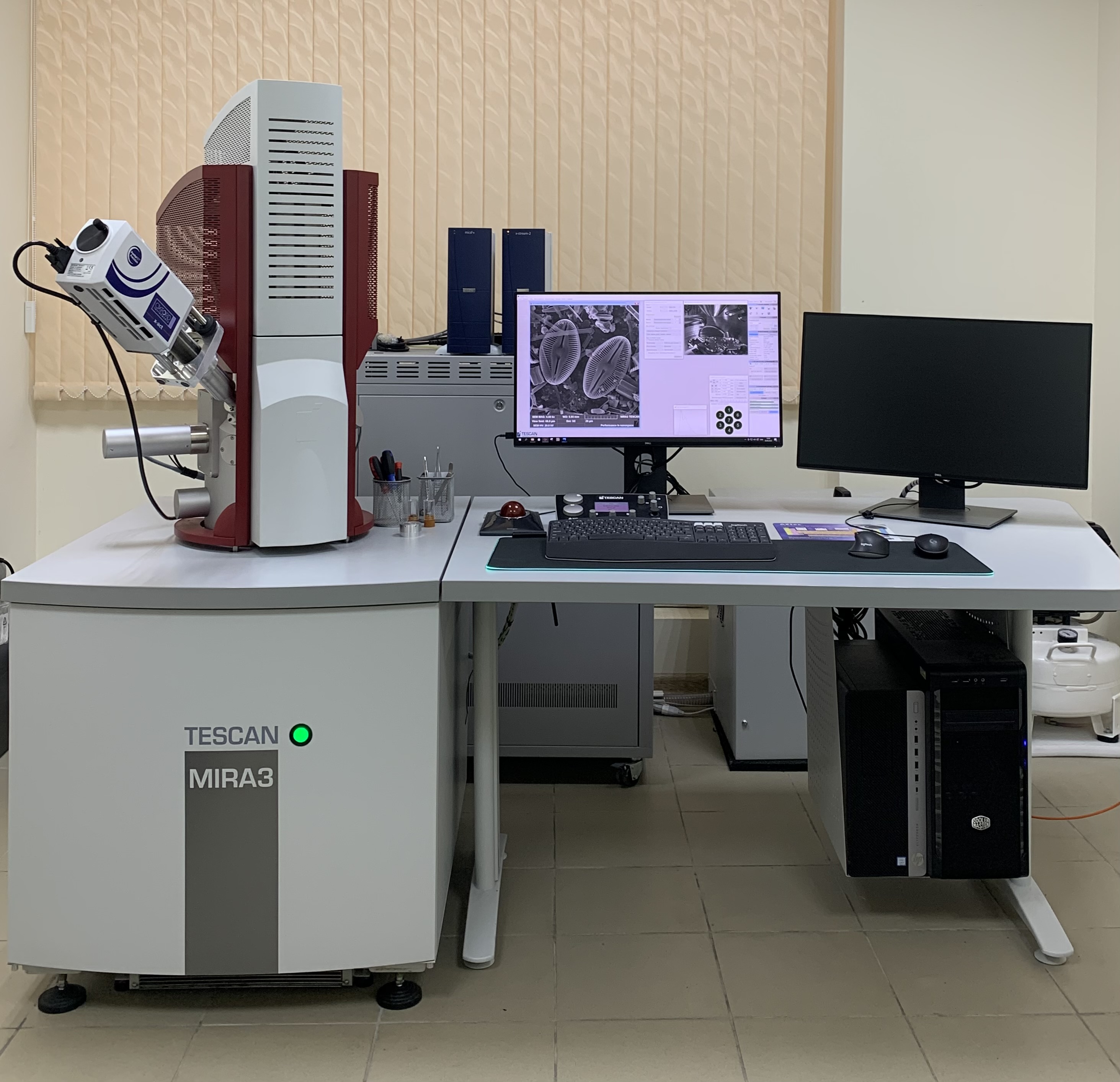
Specifications of TESCAN MIRA 3 LMH
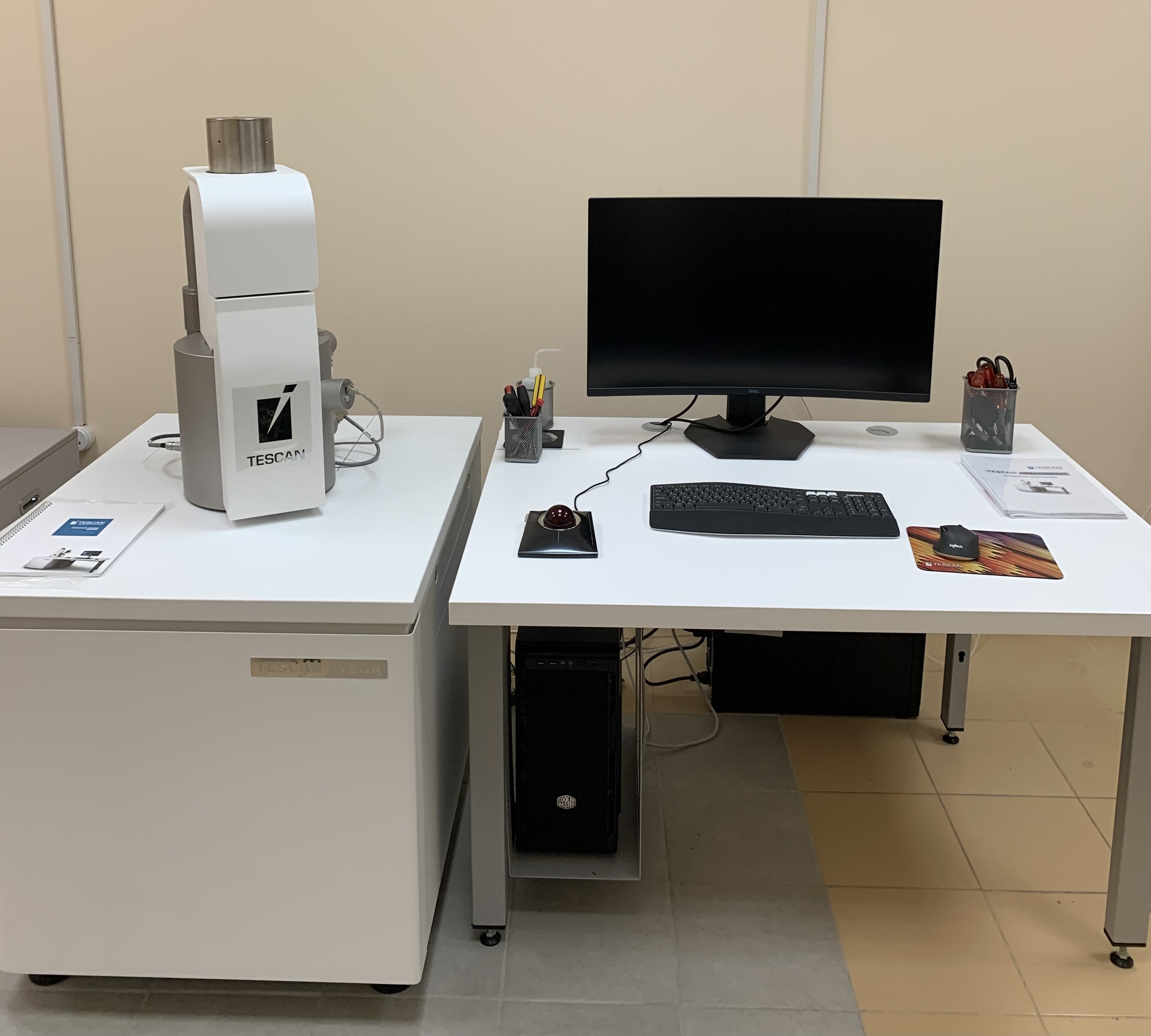
- TESCAN VEGA COMPACT LMH (TESCAN, Czech Republic), equipped with a tungsten thermionic-emission cathode, modern electron optics based on the unique four-lens Wide Field Optics™ technology with a patented intermediate lens, and secondary and backscattered electron detectors. Spatial resolution of 3.0 nm at an accelerating voltage of up to 30 kV. Commissioned in 2022;
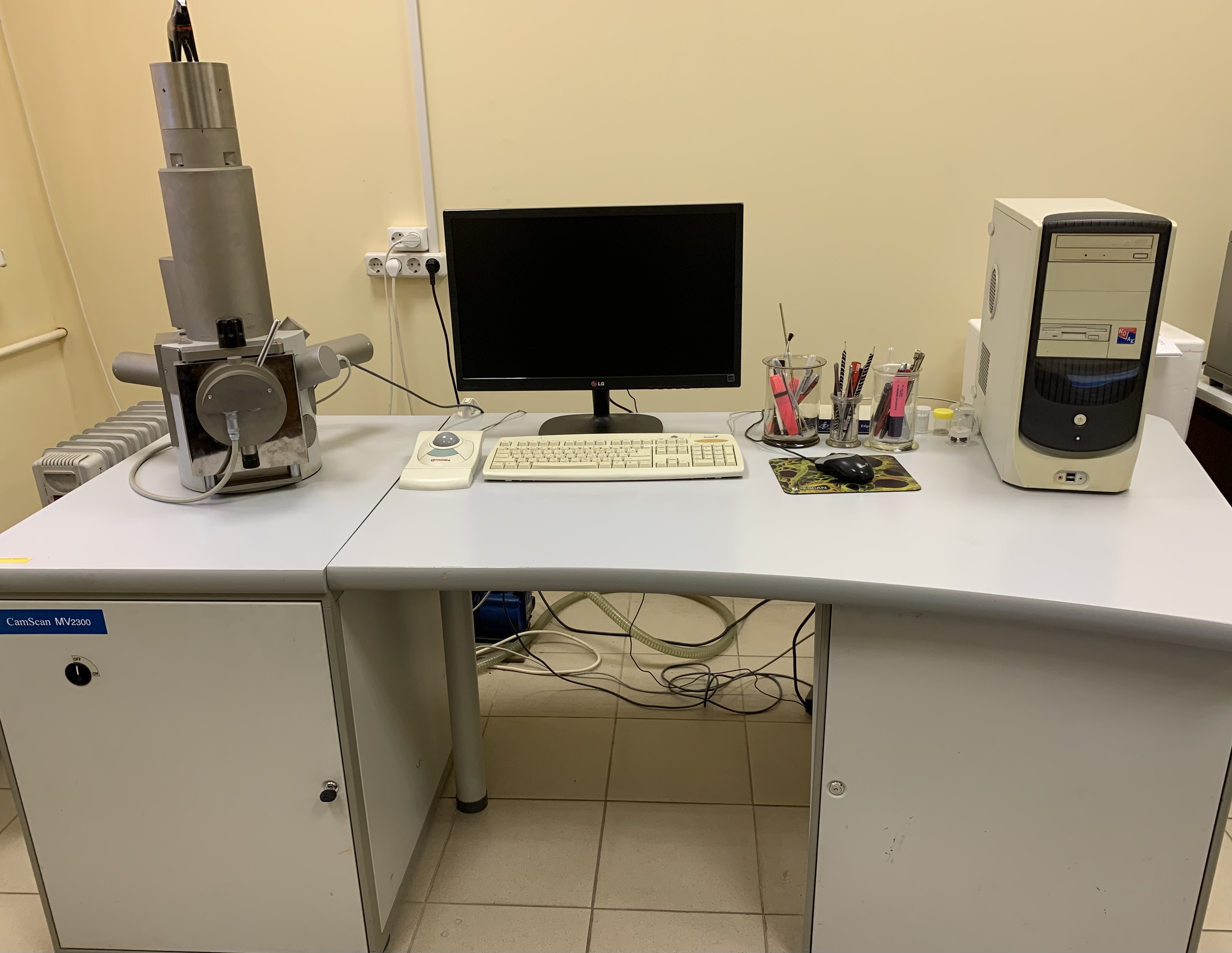
- TESCAN Vega TS5130MM (TESCAN, Czech Republic), with a tungsten cathode and secondary and backscattered electron detectors. Accelerating voltage up to 30 kV. Resolution for biological objects of about 15 nm. Commissioned in 2002.
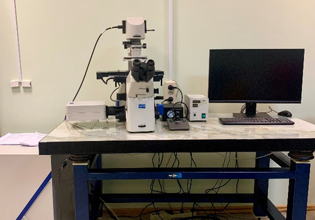
- Nexcope NCF950 inverted confocal biological microscope (China), for brightfield and multichannel fluorescence imaging with 4-channel light sources and detectors (405, 488, 561, 540 nm), capable of real-time high-resolution imaging. Scanning modes: X-Y, X-Y-Z, X-Y-T. 12-bit image depth. Commissioned in 2024.
- KEYENCE VHX-1000 research video microscope (Japan) — a versatile digital microscope with various telecentric zoom lenses providing magnification ranges of 100×–1000×, 250×–2500×, and 500×–5000×. Designed for analytical research with automatic 3D imaging and real-time 2D and 3D measurements. Allows the study of the morphology of biological objects and the nature, localization and mechanisms of cellular damage resulting from exposure to various chemicals or pollutants. Capable of detecting taxonomically significant characters in aquatic organisms or characters that change in invasive species in habitats new to them.
Critical point drying units:
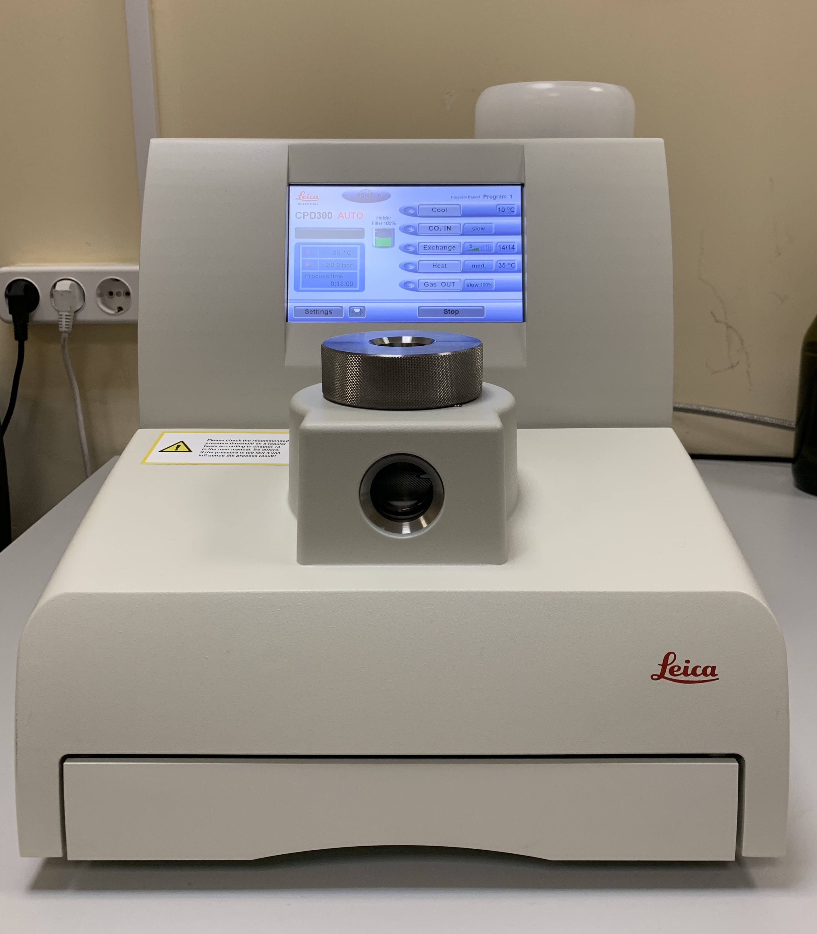
- Leica EM CPD300 (Leica Microsystems Vertrieb GmbH, Austria) — a system for automatic gentle critical-point drying of biological and industrial samples for study by scanning electron microscopy under high-vacuum conditions with maximum preservation of their morphology. Drying of biological samples pre-dehydrated through a graded series of ethanol and acetone concentrations. Commissioned in 2020;
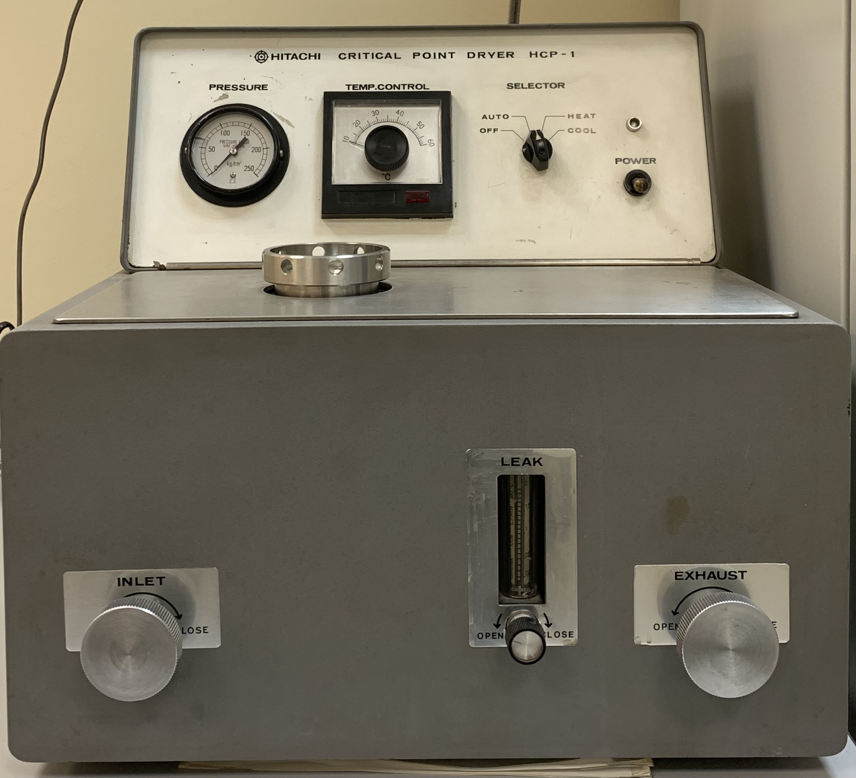
- Hitachi Critical Point Dryer HCP-1 (Hitachi Ltd., Japan) — a system for automatic gentle critical-point drying of biological and industrial samples for study by scanning electron microscopy under high-vacuum conditions with maximum preservation of their morphology; drying of biological samples pre-dehydrated through a graded series of ethanol and acetone concentrations. Commissioned in 1975.
Sputter coating units:
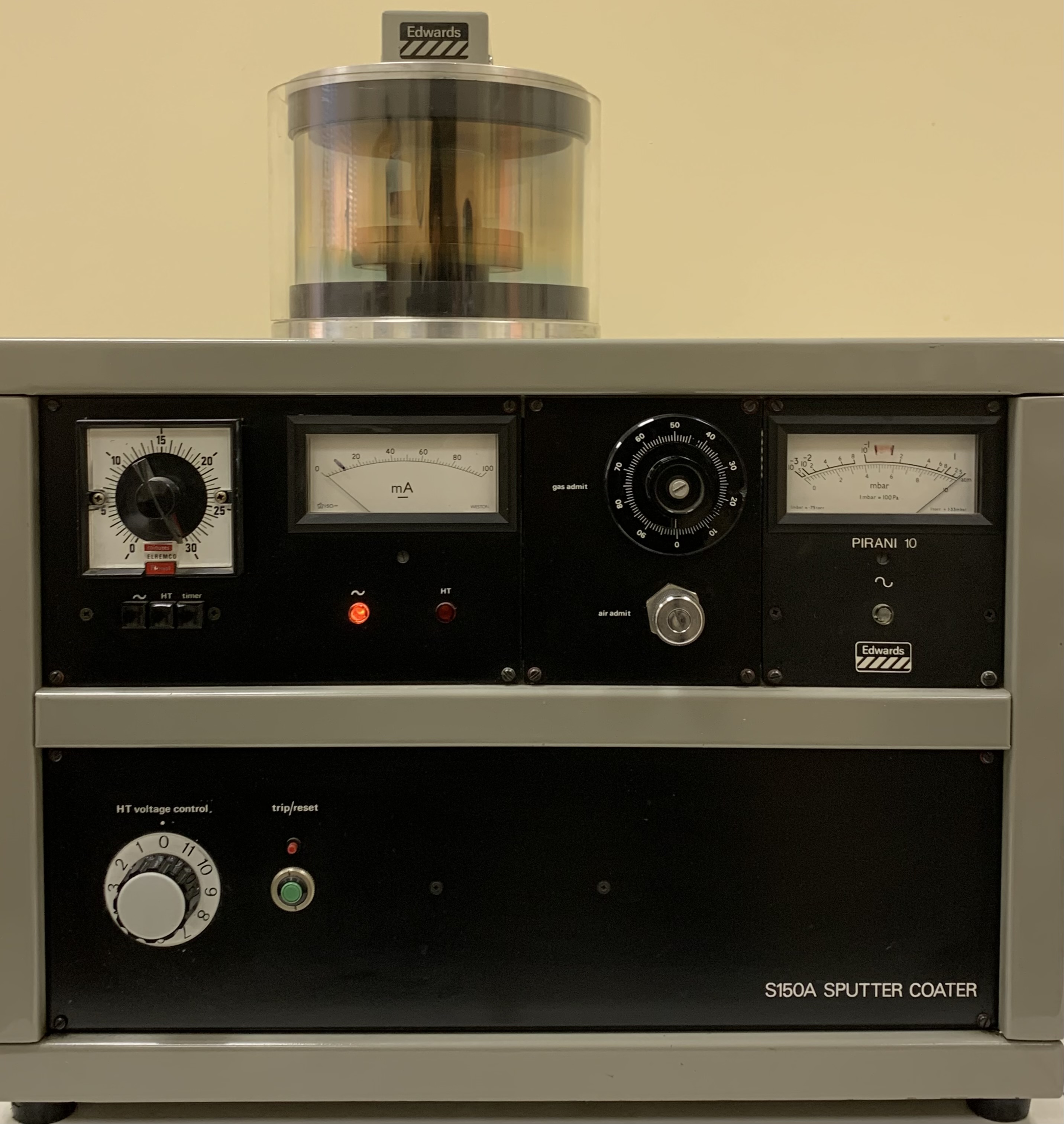
- S150A Sputter Coater (Quorum Technologies Ltd., UK) — application of a thin gold or carbon coating to samples under medium-vacuum conditions to produce a conductive surface for SEM studies. Commissioned in 2020.
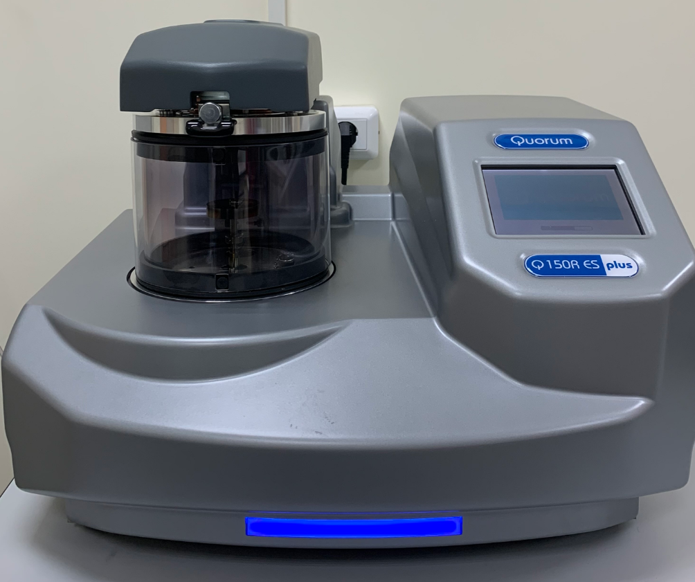
- Q150R ES Plus (Quorum Technologies Ltd, United Kingdom) — a sample preparation device for preparing samples by vapor-phase deposition of carbon or metal onto non-conductive biological and industrial samples under medium-vacuum conditions to create a conductive surface for SEM studies. Commissioned in 1980.
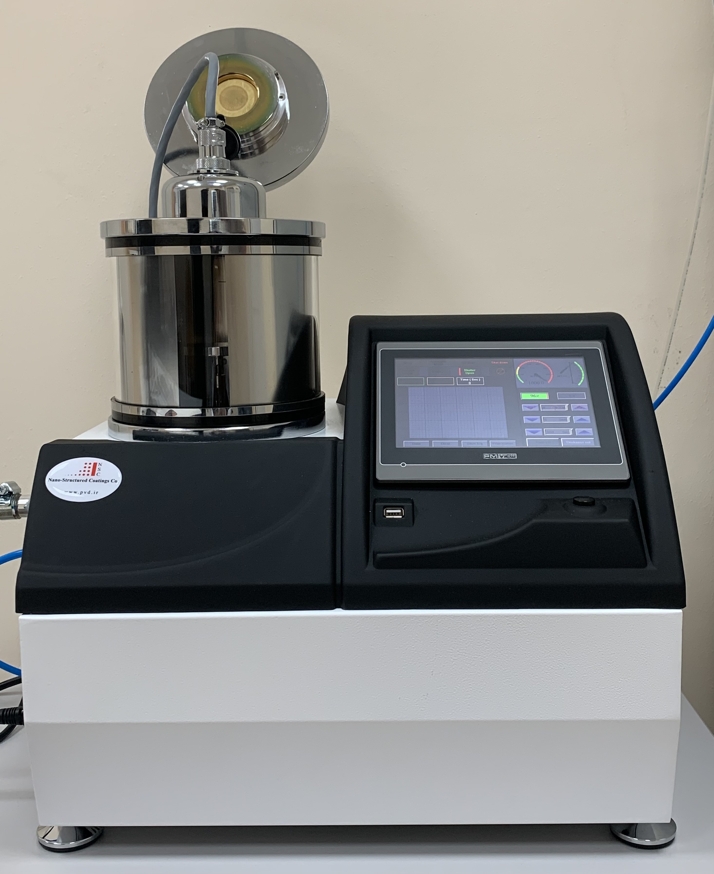
- DSCR (Nano-Structured Coatings Company, Iran) — an automatic sputter coating unit for producing carbon and metal coatings for SEM studies. Commissioned in 2023.
Light optical microscopes:
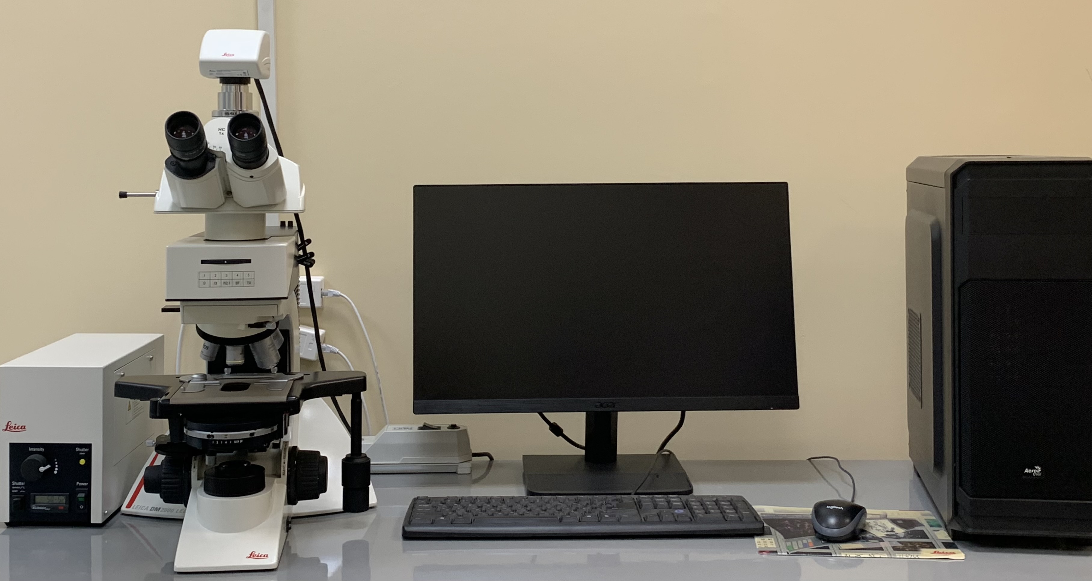
- LEICA DM2000M upright microscope (Leica Microsystems, Vertrieb GmbH, China), equipped with a Leica DMC4500 digital camera — a modern optical microscope for studying biological objects in transmitted light by brightfield, darkfield, differential interference contrast, and fluorescence methods, with a basic optical magnification of 50×–1000×, for preparing micrographs and morphometry. Commissioned in 2020;
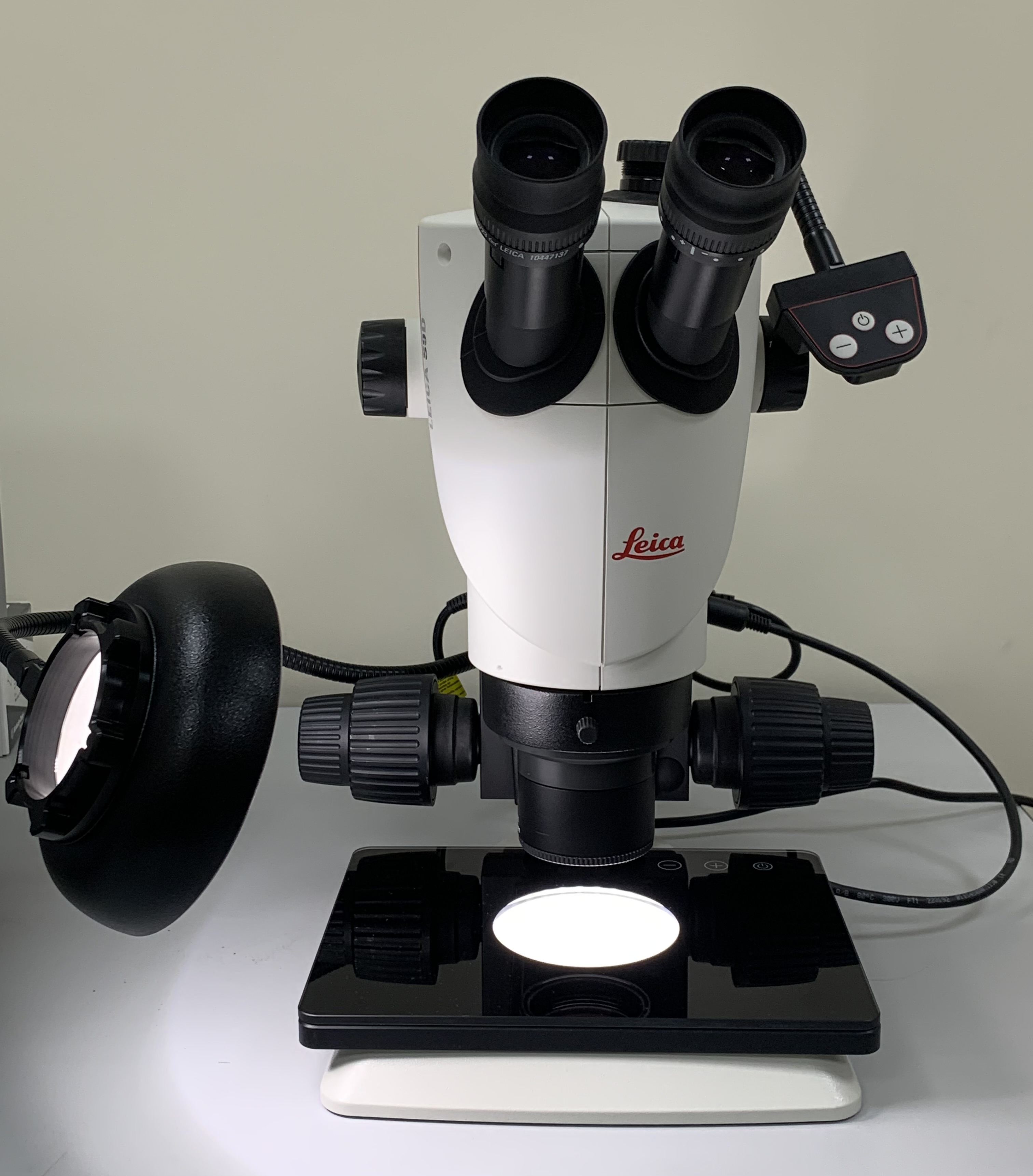
- LEICA S9D stereomicroscope (Leica Microsystems, Vertrieb GmbH, Singapore), equipped with a Leica DMC4500 digital camera for viewing biological objects in reflected and transmitted light. A stereomicroscope for preliminary study of the surface morphology of biological objects at low magnifications (6×–55×) and for obtaining stereoscopic images, used in preparing specimens for SEM. Commissioned in 2020;
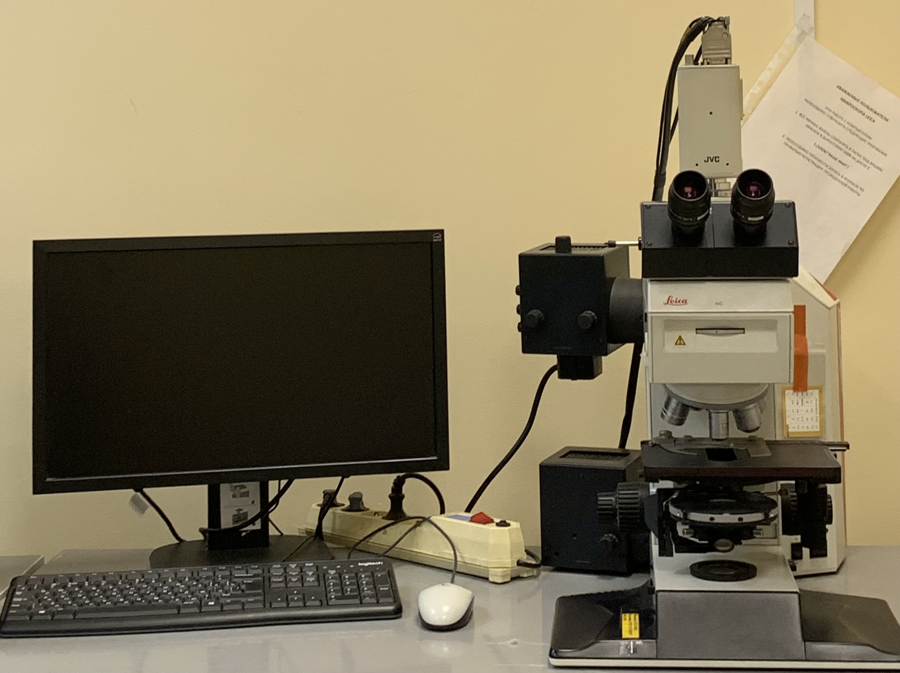
- LEICA DMR (Leica Microsystems, Germany), equipped with a JVC 3-CCD C-mount camera, for studying biological objects in transmitted light by brightfield and darkfield methods, for preparing micrographs, and for morphometry. Commissioned in 2003.