

Results
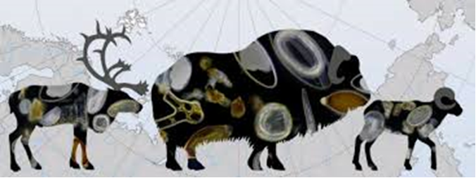
1. The biological diversity of parasitic worms in Arctic ungulates was studied: domestic and wild reindeer, muskox, and snow sheep. This first study in 60 years using modern methods led to the discovery of parasites not previously recorded in the Palearctic. One hypothesis under consideration is that they may have been introduced from North America along with muskoxen brought into the country in the 1970s. The information obtained is not only of fundamental value but also serves as a basis for developing an anti-parasitic action plan for reindeer husbandry (Food Security Doctrine) and for rare-species conservation programs (the «Ecology» National Project). This is important both for the indigenous small-numbered peoples of the North and consumers of reindeer husbandry products, and for the welfare of ungulates in nature reserves.
Link to the publication: https://doi.org/10.3390/d15050672
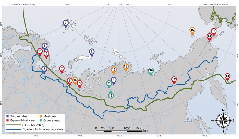
Publication: Diversity and Distribution of Helminths in Wild Ruminants of the Russian Arctic: Reindeer (Rangifer tarandus), Muskoxen (Ovibos moschatus), and Snow Sheep (Ovis nivicola) / O. A. Loginova, S. B. Rozenfeld; T. P. Sipko, I. A. Mizin, D. V. Panchenko, K. A. Laishev, M. G. Bondar, L. A. Kolpashchikov, A. R. Gruzdev, P. S. Kulemeev, D. I. Litovka, M. N. Semerikova, V. N. Mamontov, E. G. Mamaev, S. E. Spiridonov // Diversity. – 2023. – № 15. – p. 672.
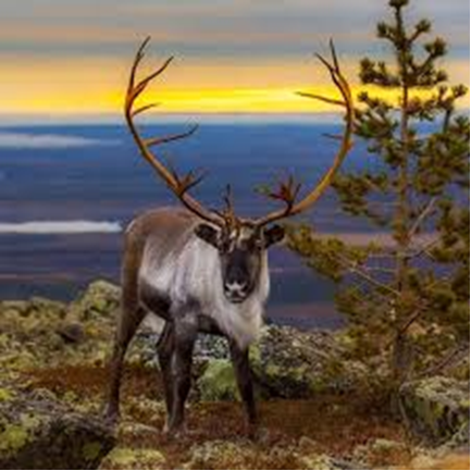
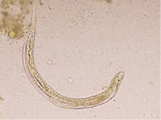
2. Researchers from the Center of Parasitology of IEE RAS, together with specialists from the «Joint Directorate of Taymyr Nature Reserves,» studied the feces of wild reindeer (Rangifer tarandus) to determine which helminths parasitize them. In addition to gastrointestinal parasitic nematodes of the order Strongylida (found as eggs and live larvae), characteristic of all ruminants, and trematodes of the genus Paramphistomum (eggs), typical of reindeer, larvae of nematodes of the family Protostrongylidae belonging to the species Varestrongylus eleguneniensis and Orthostrongylus macrotis. Both species were recorded in the Palearctic for the first time, and the latter was found in reindeer for the first time. These helminths parasitize the respiratory system and could potentially pose a threat to the Taymyr population.
Published: Loginova, O.A., Kolpashchikov, L.A. & Spiridonov, S.E. First report of Orthostrongylus sp. (Nematoda: Protostrongylidae) in wild reindeer (Rangifer tarandus) from the Taimyr,
https://doi.org/10.1007/s00436-022-07754-7
3. It was shown that the mitochondrial genomes of hairworms (Nematomorpha) are enriched with long inverted repeats embedded within the coding sequences of their genes. This is a highly unusual feature of hairworms, so far noted only for organisms of this phylum in the Animal Kingdom. Repeats in the mitochondrial genome of the hairworm Parachordodes pustulosus were obtained and annotated. Using genomic and transcriptomic libraries, the effect of inverted repeats on mitochondrial genome coverage was investigated. Pronounced drops in read coverage coincide with regions containing long inverted repeats, marking «blind spots» of short-read DNA and RNA libraries. Phylogenetic inference based on the new data reveals numerous discrepancies between the traditional Nematomorpha classification system and the molecular data.
Nikolaeva O.V., Beregova A.M., Efeykin B.D., Miroliubova T.S., Zhuravlev A.Yu., IvantsovA.Yu., Mikhailov K.V., Spiridonov S.E., Aleoshin V.V., 2023. Expression of Hairpin-Enriched Mitochondrial DNA in Two Hairworm Species (Nematomorpha) // International Journal of Molecular Sciences. V. 24. №14:11411. https://doi.org/10.3390/ijms241411411.
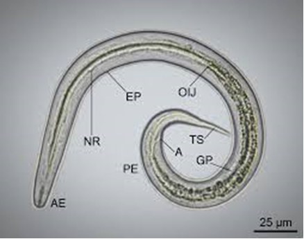
4. Patent No. 2787526, Russian Federation, IPC A61B 10/00 (2006.01), CPC A61B 10/00 (2022.08), was obtained: A method of topographic orientation for detecting the nerve ring in larvae of parasitic nematodes of the order Strongylida: No. 2022111459: filed 27.04.2022: published 10.01.2023 / O.A. Loginova; applicant IEE RAS — 11 pp.: ill.
5. The first molecular phylogenetic study of gregarines specific to centipedes — families Dactylophoridae and Trichorhynchidae — was carried out. It was reliably shown that these two groups together represent a distinct early evolutionary lineage of terrestrial gregarines, and a diagnosis for a new superfamily, Dactylophoroidea (Fig. 1). In addition, a new species for the genus Trichorhynchus — Trichorhynchus efeykini sp. n. (Fig. 1), the second species for the entire family and genus, and the first to be described in the last 140 years.
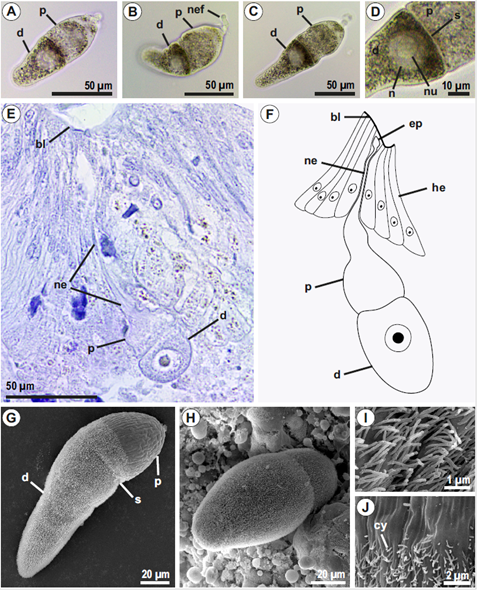
Published: Tatiana S. Miroliubova, Kirill V. Mikhailov, Timur G. Simdyanov, Vladimir V. Aleoshin,
6. Laboratory staff studied the common liver fluke (Fasciola hepatica). This parasitic worm, which lives in the liver of herbivores and humans, is known on every inhabited continent. Up to 180 million people and more than 700 million animals are at risk of infection each year. The main method of in-vivo diagnosis of fascioliasis is the detection of eggs in feces. One might expect that the appearance of the egg would have been studied in full detail over the past centuries. However, it turned out that this was not the case. In some Fasciola eggs, «tails» were found that had previously been thought to occur only in the giant liver fluke (Fascioloides magna). The descriptions and images obtained provide a detailed picture of the structure of Fasciola eggs and should help physicians and veterinarians worldwide in diagnosing fascioliasis. Link to the publication: https://doi.org/10.1016/j.fawpar.2024.e00237
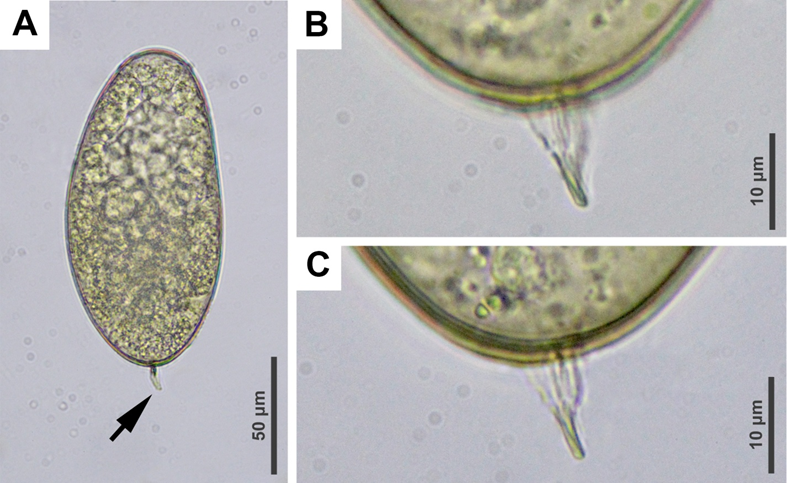
Publication: Updates on egg morphology, host range, and distribution / O. Loginova, B. Efeykin, A. Krutikova, I. Mizin, S. Spiridonov // Food and Waterborne Parasitology. – 2024. – №