On November 21, a practical lesson was held at the IEE RAS, based at the Electron Microscopy Room, as part of the Competition for schoolchildren of specialized classes, under the guidance of Anna Neretina, PhD in Biology, research fellow at the A.N. Severtsov Institute of Ecology and Evolution of the Russian Academy of Sciences.
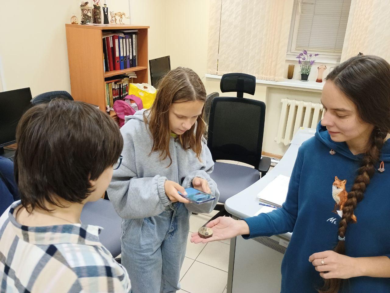
The children became familiar with the devices used to prepare microscopic hydrobionts for examination using scanning electron microscopy methods, and the principles of their operation (Fig. 1).

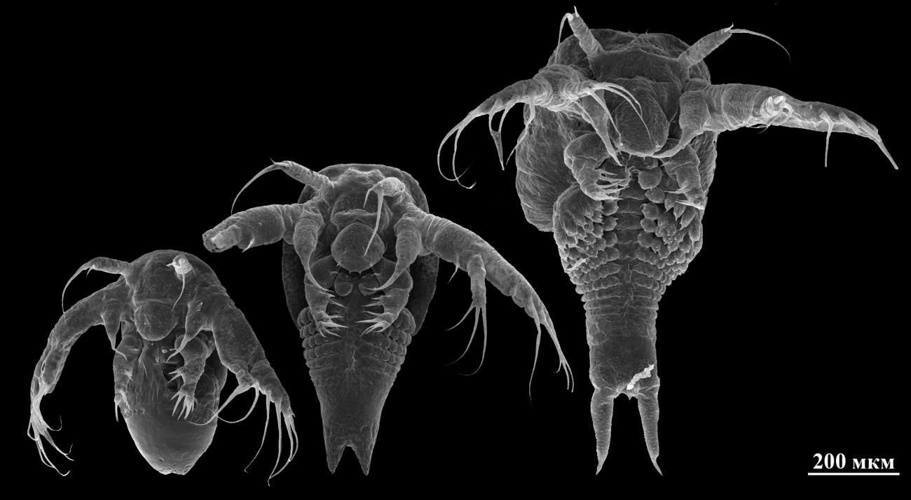
The excursionists were especially delighted by the larvae of the summer shieldfish (Triops cancriformis), hatched from dormant eggs by Denis Polyakov, a 4th-year student of the Department of Invertebrate Zoology at Lomonosov Moscow State University (Fig. 2).
The beauty and unpretentiousness of shieldfish to cultivation conditions contributed to the fact that this group is very popular not only with biologists and professional aquarists, but also with children. The participants of the lesson were among the first to see what the larvae of Triops cancriformis look like.

