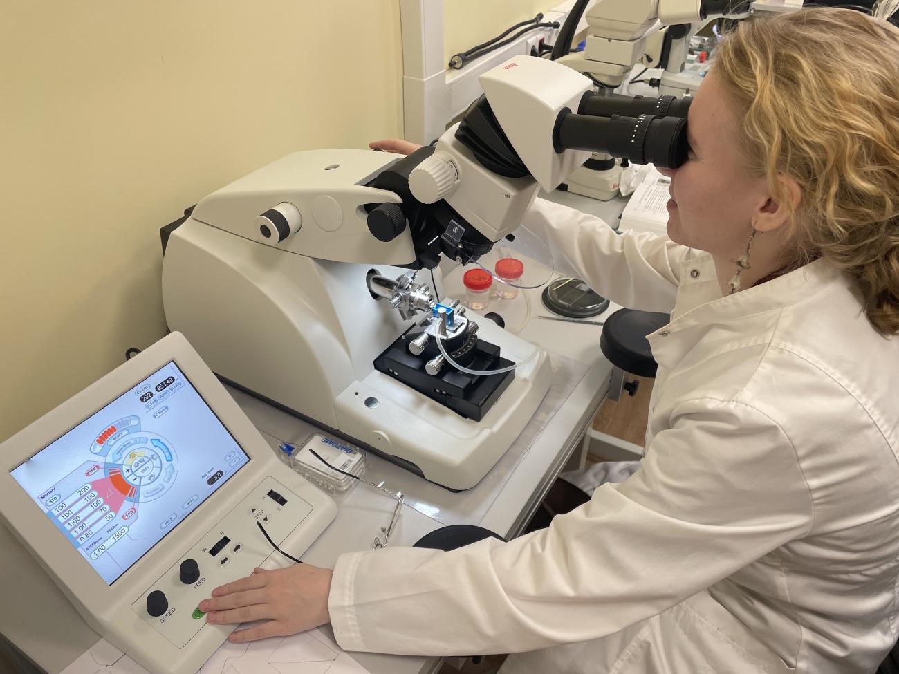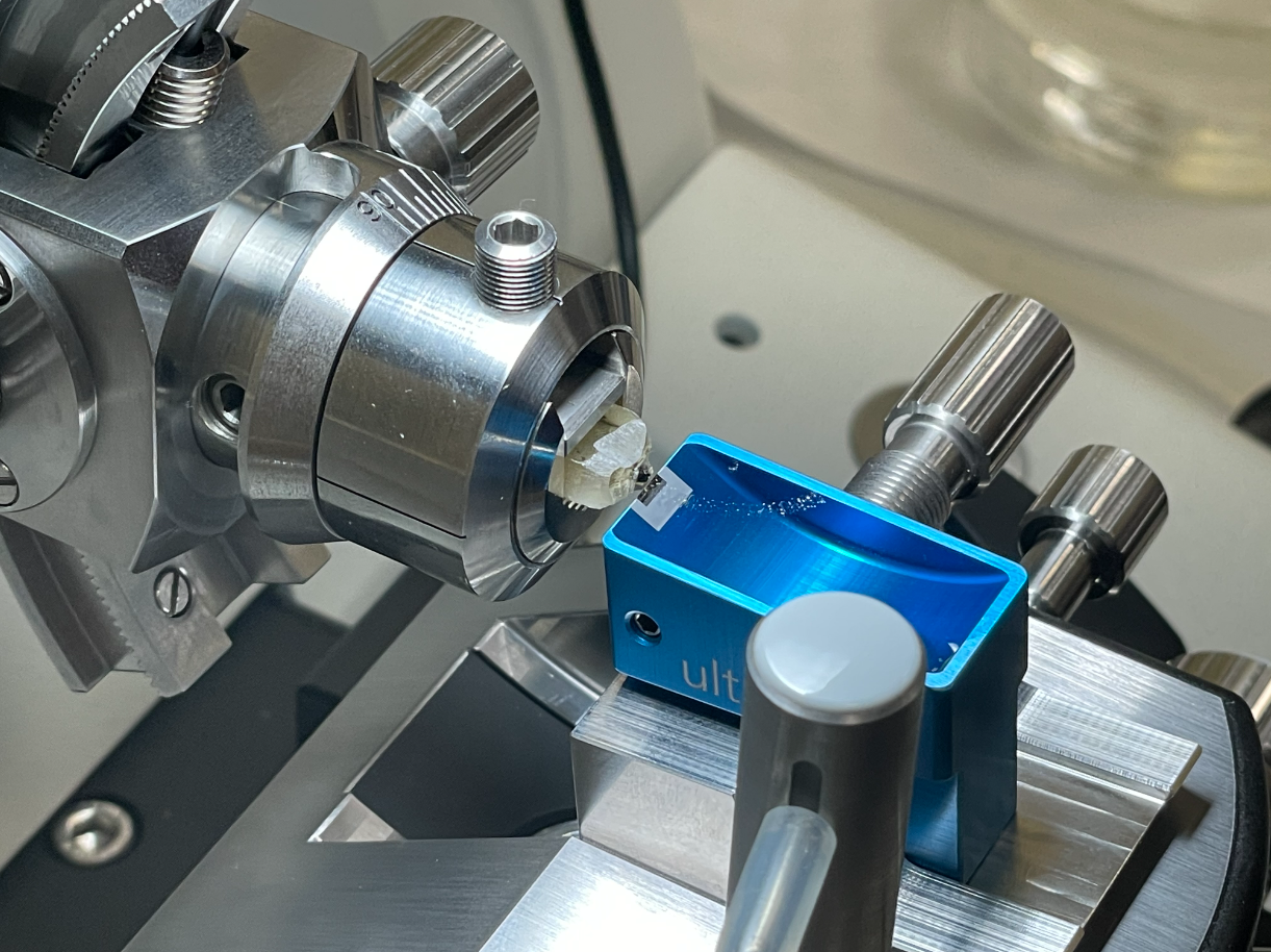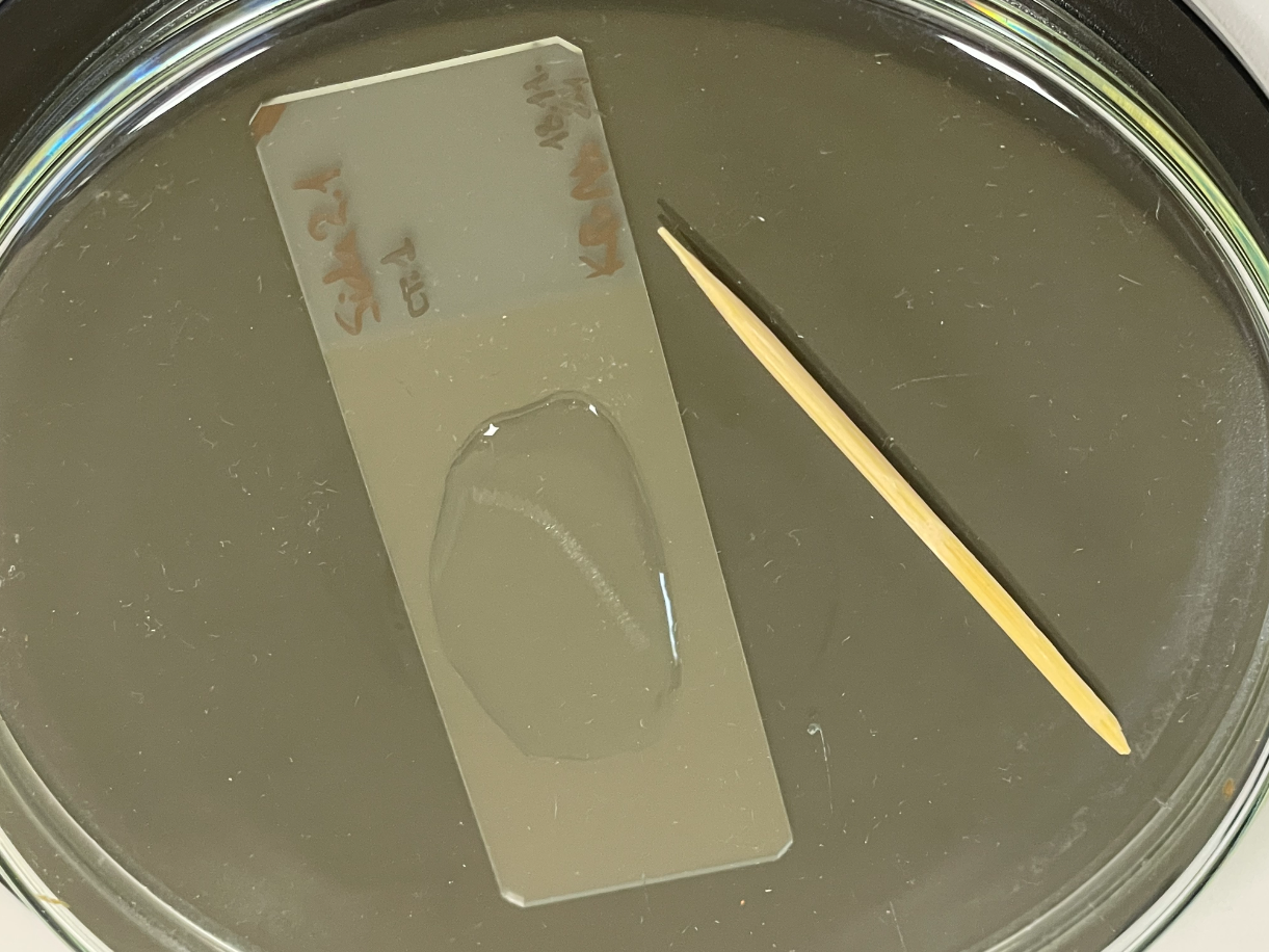
We are happy to share a joyful event with you. This year, Varvara Krolenko, a true master of making sections of biological objects, entered the graduate school of Alexey Alekseevich Kotov. Under the program for updating the equipment base of organizations of the Ministry of Science and Higher Education of the Russian Federation, the institute purchased a Leica EM UC7 ultratome and today, with joint efforts, we launched it. The device is designed to obtain semi-thin and ultra-thin sections of objects embedded in epoxy resins. Varya will use it to cut water fleas (Crustacea: Cladocera) for her PhD dissertation.

After sectioning, Varvara examines the sections using a transmission electron microscope and, based on the data obtained, will perform volumetric reconstructions of the organ systems of cladocerans.
The appearance of such a device at the institute opens the way to research that is at the forefront of science. Example.

But sectioning biological objects is only one of the preparatory stages for conducting ultrastructural studies. In order to fully perform such work in our institute, it is necessary to purchase a transmission electron microscope, as well as equip a separate room for sample preparation. Now in the room where the ultratome is registered, 4 people, 3 direct microscopes, 4 stereomicroscopes and many other valuable things fit on 18 square meters. In addition, schoolchildren and students from other organizations come to visit us on a regular basis. We all have very different tasks, from routine faunistics and floristry to boring taxonomy and even work with living objects. Expanding the list of laboratory topics, we also dream of increasing the areas for work. And we definitely have enough imagination and strength to master and equip them.
