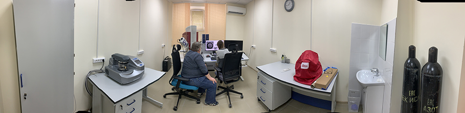
A.N. Severtsov Institute of Ecology and Evolution of Russian Academy of Sciences within the framework of the Ministry of Education and Science project "Updating the instrument base of the leading organizations performing research and development in the academic sector of science" has acquired new equipment for the electron microscopy room:
- Scanning electron microscope TESCAN MIRA 3 LMH
- is equipped with a Schottky cathode, a detector for studying samples "in the light" and an energy dispersive microanalysis system AZtecOneX-act. (SEM TESCAN MIRA 3 LMH is one of the most modern hi-tech devices - a universal analytical complex for studying the morphology and ultrastructure of biological and industrial objects with an ultrahigh spatial resolution based on a scanning electron microscope and conducting a semi-quantitative microanalysis of biological samples. The Schottky auto-emission cathode provides resolution - 1.2 nm at 30 kV, magnification range without distortion of the field of view from 2 X to 1 000 000 X, scanning speed 20 ns / pixel, image saving up to 16 384 X 6 384 pixels and high performance. Energy dispersive microanalysis system AZtecOneX-act allows obtaining SEM images in secondary or reflected electrons, building maps of the distribution of elements in a certain area and obtaining color representations of several elements in one summary image with the possibility of overlaying them on an electronic image).
- Sputtering installation Q150R ES Plus (Quorum Technologies)
(Sample preparation device for rapid preparation of samples by deposition of carbon or metal from the vapor phase on non-conductive biological and industrial samples to create a conductive surface).
- Automatic drying at a critical point Leica EM CPD300
(A system for automatic soft drying of biological and industrial samples at a critical point for examination in a scanning electron microscope in high vacuum conditions. Drying of such samples as pollen, plant and animal tissues with the maximum possible preservation of their morphology is ensured).
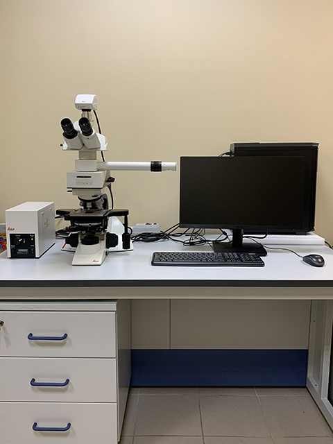
- Stereo microscope Leica S9D
(Stereomicroscope for preliminary examination of the surface morphology of biological objects at low magnifications (from 6 to 55 X) and obtaining a stereoscopic image, for making preparations for SEM).
- Straight microscope Leica DM 2000M with drawing device
(Optical microscope for studying biological objects in transmitted light through the methods of bright and dark fields, differential interference contrast, fluorescence with a base optical magnification from 50 X to 1000 X, preparation of micrographs and morphometry).
On July 8, 2020, the ceremonial start of the electron microscope took place in the electron microscopy room with cutting the ribbon by the director of the institute, academician V.V. Rozhnov.